Anatomy of the eye coloring page
Anatomy of the eye coloring page – free to print on A4 or US Letter, to download as a PDF or to color online.
Tip: turn off "Headers and footers" in the print window.
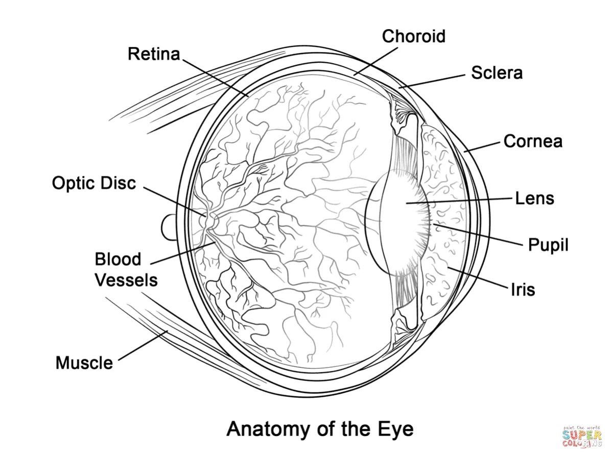
This educational drawing shows a cross-section of the human eye with its main parts labeled: the cornea, iris, pupil, lens, retina, choroid, sclera, optic disc, blood vessels and the muscles that move the eye. It suits older children and teens, roughly ages 10 to 12 and up, who are learning about the senses in school. The labels help them connect each part to its job, so it works well as a study aid.
For a clear result, color the iris and the lens in lighter, distinct shades so they stand apart from the retina, and use a fine pen or pencil for the blood vessels to keep the detail readable.
Ideas for the classroom
- Have students color each labeled part and write one sentence explaining its function.
- Pair the page with a lesson on how light travels through the cornea and lens to the retina.
Printing tips
Choose A4 if you are in Europe and US Letter in the US or Canada, so nothing gets cut off. Plain 80 g paper works for crayons; use thicker paper for markers or watercolors.