Human cell diagram to color and label coloring page
Human cell diagram to color and label coloring page – free to print on A4 or US Letter, to download as a PDF or to color online.
Tip: turn off "Headers and footers" in the print window.
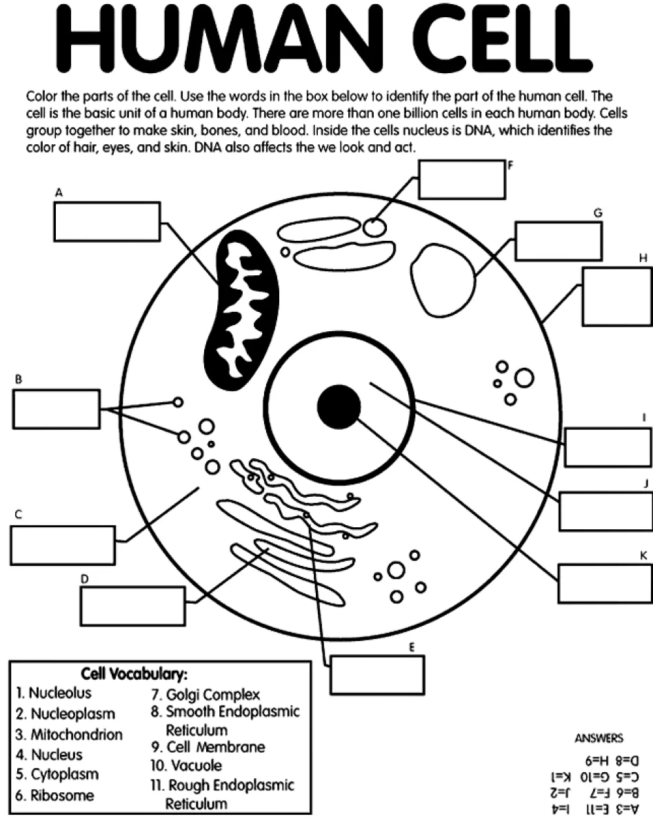
This educational drawing shows a large human cell with its main parts outlined: the nucleus with its nucleolus, the cell membrane, the mitochondrion, the Golgi complex, the endoplasmic reticulum, ribosomes, vacuoles and cytoplasm. Blank label boxes are placed around the cell, and a vocabulary list in the corner gives the names to match. It suits older children, roughly ages 10 to 12, who are learning cell biology in school.
Tip: color each organelle a different bright shade and use the same color for its label box, so the parts are easy to tell apart at a glance.
Ideas for the classroom
- Have students color each organelle, then write its function in the matching label box.
- Use the page as a quiz: students fill in the labels from memory before checking the vocabulary list.
- Ask pairs to explain to each other what the nucleus and mitochondria do in the cell.
Printing tips
Choose A4 if you are in Europe and US Letter in the US or Canada, so nothing gets cut off. Plain 80 g paper works for crayons; use thicker paper for markers or watercolors.